Zebrafish Embryo Gets The Google Earth Treatment
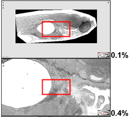
Electron microscopes are amazing tools for getting detailed closeups of the things inside a cell that make it go, but when it comes to putting those things in the context of a whole organism, they’re not such a great tool. A team at the University of Leden in the Netherlands is helping microscope users focus on the bigger picture. They’ve created a nearly complete atlas of a zebrafish embryo that researchers can navigate by zooming in and out through various levels of detail — just like Google Earth, except with the inside of a baby fish.
Using a technique called virtual nanoscopy, the team stitched together tens of thousands of electron microscope images provided by researchers around the world. The resulting mosaic is a 281 gigapixel behemoth with a resolution equivalent to 16 million dpi, crafted from over 26,000 individual images. With images from multiple levels of magnification, researchers will be able to take a look at everything from the entire 1.5mm embryo to its most basic, nanometer-long components. The whole thing will be available to both cell biologists and anyone who is just curious what a baby zebrafish looks like from the inside using the Journal of Cell Biology’s Dataviewer tool.
Now, zooming in on the features of individual cells and getting a better look at how they interact is as easy as finding out how big your neighbor’s pool is. Just place your cursor and keep zooming until you start feeling like a voyeur. So for those of you who have ever wished you could take a leisurely stroll around the cellular structure of a developing zebrafish embryo — and let’s face it, who among us hasn’t fantasized about that — your day is quickly approaching.
(via ScienceDaily)
- Because it bears saying again: Electron microscopes are awesome
- Though zebrafish embryos will probably not inspire any Persian Rug Designs
Have a tip we should know? tips@themarysue.com
